
examination of cranial nerves pdf
Cranial nerve examination is a fundamental component of neurological assessment‚ providing critical insights into diagnosing neurological conditions and identifying abnormalities in cranial nerve function.
1.1 Importance of Cranial Nerve Examination in Neurology
Cranial nerve examination is a cornerstone in neurology‚ enabling the identification of neurological deficits and localization of brainstem or peripheral nerve lesions. It aids in diagnosing conditions like strokes‚ tumors‚ or neuropathies by assessing sensory‚ motor‚ and autonomic functions. A thorough examination helps guide further investigations and treatment‚ ensuring timely intervention for conditions affecting vision‚ hearing‚ swallowing‚ or facial movements‚ ultimately improving patient outcomes and quality of life.
1.2 Overview of the 12 Cranial Nerves
The 12 cranial nerves originate from the brainstem and control essential functions. They include the olfactory (I)‚ optic (II)‚ oculomotor (III)‚ trochlear (IV)‚ trigeminal (V)‚ abducens (VI)‚ facial (VII)‚ vestibulocochlear (VIII)‚ glossopharyngeal (IX)‚ vagus (X)‚ accessory (XI)‚ and hypoglossal (XII) nerves. These nerves manage sensory‚ motor‚ and autonomic functions‚ such as vision‚ hearing‚ smell‚ taste‚ swallowing‚ and muscle control‚ playing a vital role in maintaining bodily functions and overall health.
Equipment and Preparation for Cranial Nerve Examination
Essential tools include a Snellen chart‚ penlight‚ otoscope‚ and tongue depressor. Ensure proper patient positioning and introduce yourself to establish comfort and understanding of the examination process.
2.1 Essential Tools for the Examination
The examination requires a Snellen chart for visual acuity‚ a penlight for pupil reactions‚ an otoscope for hearing assessment‚ and a tongue depressor for evaluating tongue movements. Additional tools include a soft brush for olfactory testing and a reflex hammer for cranial nerve reflexes. Ensure all equipment is readily available to streamline the process and maintain patient comfort throughout the evaluation.
2.2 Patient Preparation and Positioning
Patient preparation involves ensuring a comfortable and well-lit environment. Position the patient sitting upright with head unsupported for optimal cranial nerve assessment. Remove any distractions and ensure the patient is relaxed. For specific tests‚ such as olfactory nerve evaluation‚ avoid strong odors. Proper positioning facilitates accurate testing of eye movements‚ facial expressions‚ and tongue protrusion‚ ensuring reliable clinical findings during the examination.
Examination of Cranial Nerves I-IV
This section focuses on assessing cranial nerves I (olfactory)‚ II (optic)‚ III (oculomotor)‚ and IV (trochlear)‚ covering tests for smell‚ vision‚ eye movements‚ and pupillary reflexes.
3.1 Olfactory Nerve (I) ⸺ Sense of Smell
The olfactory nerve (I) is assessed by testing the patient’s ability to detect and identify smells using pungent‚ non-irritating odors. Each nostril is examined separately‚ with the contralateral nostril occluded. The patient should identify the odor and its intensity. Anosmia or reduced olfaction may indicate nerve dysfunction. This test is crucial for diagnosing conditions affecting the olfactory pathway‚ providing insights into potential neurological disorders.
3.2 Optic Nerve (II) ⎻ Vision and Pupil Reaction
Examination of the optic nerve (II) involves assessing visual acuity using a Snellen chart‚ testing visual fields‚ and evaluating pupil reactions. The consensual light reflex and accommodation reflex are checked. An ophthalmoscope is used to inspect the optic disc for signs of atrophy or swelling. Abnormalities‚ such as an afferent pupillary defect‚ may indicate optic neuritis or other conditions. This evaluation is crucial for identifying optic nerve dysfunction.
3.3 Oculomotor Nerve (III) ⸺ Eye Movement and Pupillary Reflex
The oculomotor nerve (III) controls eye movement‚ eyelid elevation‚ and pupillary reflexes. Examination involves assessing extraocular muscle function by testing eye movements in all directions. Pupillary light reflex and accommodation reflex are evaluated. Ptosis or diplopia may indicate dysfunction. The “cover-uncover” test can detect subtle deficits. Abnormal findings‚ such as anisocoria or impaired reflexes‚ may suggest nerve damage‚ aneurysms‚ or increased intracranial pressure.
3.4 Trochlear Nerve (IV) ⎻ Superior Oblique Muscle Function
The trochlear nerve (IV) is the thinnest and longest cranial nerve‚ innervating the superior oblique muscle‚ which rotates the eye downward and inward. Examination involves assessing whether the patient can perform these specific eye movements. A deficit may cause difficulty looking downward‚ such as when walking stairs. The “cover-uncover” test can reveal latent strabismus‚ while a Bielschowsky head tilt test may indicate superior oblique dysfunction.
Examination of Cranial Nerves V-VIII
This section examines cranial nerves V to VIII‚ focusing on their roles in facial sensation‚ eye movement‚ facial muscle control‚ and hearing. It provides a guide for assessing these nerves.
4.1 Trigeminal Nerve (V) ⎻ Facial Sensation and Motor Function
The trigeminal nerve‚ the largest cranial nerve‚ has three branches (ophthalmic‚ maxillary‚ mandibular) and dual roles in facial sensation and motor function. Examination involves assessing facial sensation using a cotton swab‚ testing motor function by palpating muscles of mastication‚ and evaluating the corneal reflex. Clinical findings like asymmetry or muscle atrophy can indicate dysfunction‚ aiding in the diagnosis of conditions affecting this nerve.
4.2 Abducens Nerve (VI) ⎻ Lateral Eye Movement
The abducens nerve controls the lateral rectus muscle‚ responsible for outward eye movement. Examination involves assessing lateral gaze by asking the patient to follow an object with their eyes. Abnormalities‚ such as medial strabismus or inability to abduct the eye‚ suggest dysfunction. This nerve’s evaluation is crucial for diagnosing conditions like cranial nerve palsies or increased intracranial pressure.
4.3 Facial Nerve (VII) ⎻ Facial Muscle Control
The facial nerve controls facial muscles‚ taste to the anterior tongue‚ and tear production. Examination includes assessing facial muscle symmetry‚ testing taste with sweet‚ sour‚ and salty substances‚ and evaluating the stapedius reflex. Abnormalities‚ such as facial weakness or impaired taste‚ may indicate dysfunction‚ often seen in conditions like Bell’s palsy. Accurate assessment aids in diagnosing facial nerve palsies and their underlying causes.
4.4 Vestibulocochlear Nerve (VIII) ⸺ Hearing and Balance
The vestibulocochlear nerve manages hearing and balance. Examination involves assessing hearing via whisper tests‚ tuning fork tests‚ and checking for nystagmus during caloric reflex testing. Abnormalities like hearing loss or vertigo may indicate vestibulocochlear dysfunction‚ often due to conditions such as Ménière’s disease or acoustic neuromas. Accurate evaluation is essential for diagnosing disorders affecting hearing and equilibrium;

Examination of Cranial Nerves IX-XII
Examination of cranial nerves IX-XII focuses on assessing glossopharyngeal‚ vagus‚ accessory‚ and hypoglossal nerve functions. These nerves control swallowing‚ voice‚ neck muscles‚ and tongue movements.
5.1 Glossopharyngeal Nerve (IX) ⎻ Swallowing and Taste
The glossopharyngeal nerve (IX) manages swallowing‚ taste sensation on the posterior tongue‚ and salivation. Examination involves assessing the gag reflex‚ taste perception‚ and pharyngeal muscle function. A diminished gag reflex or difficulty swallowing may indicate dysfunction. Testing taste with sweet‚ sour‚ and bitter substances helps evaluate sensory integrity. This nerve’s role in deglutition and sensory input makes it crucial for maintaining oral and pharyngeal functions.
5.2 Vagus Nerve (X) ⸺ Swallowing‚ Voice‚ and Autonomic Functions
The vagus nerve (X) is vital for various functions‚ including swallowing‚ voice modulation‚ and autonomic regulation. Assessment involves evaluating the uvular reflex‚ observing vocal cord movement‚ and checking for signs of dysphonia. Autonomic functions‚ such as heart rate variability‚ may also be tested. Dysfunction can lead to hoarseness‚ dysphagia‚ or autonomic instability. This nerve’s extensive role makes it a critical focus in neurological examinations.
5.3 Accessory Nerve (XI) ⎻ Neck and Shoulder Muscle Control
The accessory nerve (XI) controls the sternocleidomastoid and trapezius muscles‚ essential for neck rotation‚ shoulder elevation‚ and scapular stabilization. Examination involves assessing muscle strength and movement‚ such as shrugging shoulders or turning the head against resistance. Weakness may indicate nerve damage‚ leading to impaired mobility and postural issues. Accurate testing ensures proper diagnosis and treatment of related musculoskeletal and neurological conditions.
5.4 Hypoglossal Nerve (XII) ⸺ Tongue Movement
The hypoglossal nerve (XII) governs tongue protrusion‚ strength‚ and coordination. Examination involves assessing tongue movement‚ such as protrusion and lateral deviation‚ to detect weakness or atrophy. Difficulty in moving the tongue may indicate nerve damage‚ affecting speech and swallowing. Proper evaluation ensures timely identification of impairments‚ guiding appropriate interventions to restore function and improve quality of life for patients with suspected hypoglossal nerve dysfunction.
Clinical Significance of Cranial Nerve Lesions
Cranial nerve lesions are crucial in diagnosing neurological conditions‚ impacting patient outcomes. Early detection ensures timely intervention‚ improving quality of life and functional recovery.
6.1 Common Causes of Cranial Nerve Dysfunction
Cranial nerve dysfunction often results from tumors‚ aneurysms‚ infections‚ or trauma. Other causes include idiopathic conditions‚ systemic diseases‚ and vascular disorders. Early identification is crucial for effective management.
6.2 Red Flags in Cranial Nerve Examination
Red flags include sudden vision loss‚ diplopia‚ facial weakness‚ or difficulty swallowing. These symptoms may indicate serious conditions like strokes‚ tumors‚ or aneurysms. Immediate referral is crucial for prompt diagnosis and treatment.
Specialized Techniques in Cranial Nerve Assessment
Advanced diagnostic methods like EMG and imaging studies help evaluate cranial nerve function‚ aiding in precise identification of pathologies and guiding targeted interventions.
7.1 Electrodiagnostic Tests for Cranial Nerves
Electrodiagnostic tests‚ such as electromyography (EMG) and nerve conduction studies (NCS)‚ are essential for assessing cranial nerve function. These tests measure electrical activity in muscles and nerves‚ helping identify abnormalities like nerve damage or dysfunction. EMG is particularly useful for evaluating cranial nerves VII‚ X‚ XI‚ and XII‚ while NCS can assess sensory and motor functions of other cranial nerves‚ providing valuable diagnostic insights.
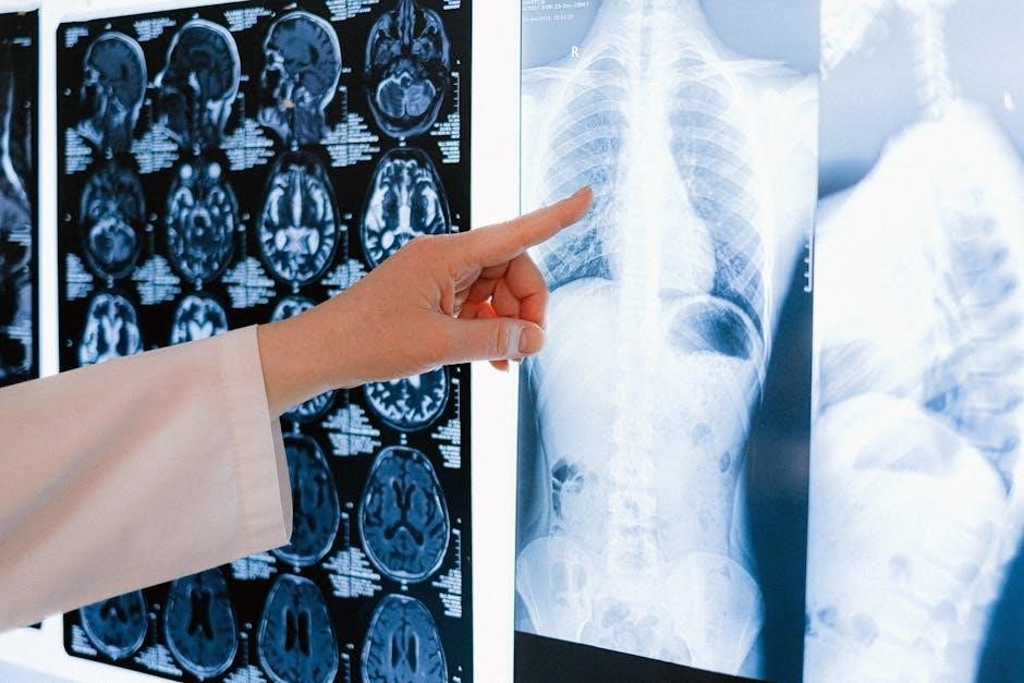
7.2 Imaging Studies for Cranial Nerve Pathologies
Imaging studies‚ such as MRI and CT scans‚ are crucial for identifying structural abnormalities affecting cranial nerves. MRI provides detailed visualization of nerve pathways‚ while CT scans are useful for detecting bony abnormalities. These modalities help diagnose conditions like tumors‚ aneurysms‚ or nerve compression‚ complementing clinical findings and guiding further management. Advanced imaging techniques ensure accurate localization of cranial nerve pathologies‚ aiding in precise diagnosis and treatment planning.
OSCE Checklist for Cranial Nerve Examination
An OSCE checklist ensures a systematic approach‚ covering equipment preparation‚ patient introduction‚ cranial nerve inspection‚ and documentation of abnormalities‚ aiding in thorough and organized assessment.
8.1 Step-by-Step Guide for Medical Students
A step-by-step guide for medical students involves preparing equipment‚ introducing oneself‚ positioning the patient‚ and systematically testing each cranial nerve. This structured approach ensures comprehensive assessment‚ beginning with visual acuity and pupil reactions for CN II‚ followed by motor function tests for CN III and IV‚ and progressing through sensory and motor evaluations for the remaining nerves. Documentation of findings is crucial for accurate diagnosis.
8.2 Common Mistakes to Avoid During the Exam
Common mistakes include inadequate patient preparation‚ poor lighting‚ and failure to systematically test each cranial nerve. Students must avoid rushing the examination‚ ensure proper patient positioning‚ and accurately document findings. Neglecting to test both eyes or missing subtle signs like anisocoria can lead to incomplete assessments. Proper equipment use and attention to detail are essential for accurate diagnosis and effective patient care.
Case Studies and Clinical Scenarios
Case studies highlight real-life applications of cranial nerve examinations‚ showcasing diagnostic challenges and uncommon presentations‚ such as bilateral cranial neuropathies or rare syndromes affecting multiple nerves simultaneously.
9.1 Examples of Cranial Nerve Palsies and Their Presentation
Cranial nerve palsies can present in isolation or as part of complex syndromes. For instance‚ bilateral involvement of trigeminal (V)‚ abducens (VI)‚ and facial (VII) nerves may result in facial weakness and impaired eye movement. A case involving Horners syndrome and lower cranial nerve palsies (IX‚ X‚ XI‚ XII) demonstrated difficulties in swallowing and speech. These examples highlight the diverse clinical manifestations and diagnostic challenges in cranial nerve disorders.
9.2 Diagnostic Challenges in Cranial Nerve Disorders
Diagnosing cranial nerve disorders presents challenges due to overlapping symptoms with other neurological conditions. Variability in clinical presentation and the need for specialized tests like EMG or imaging complicate accurate diagnosis. Additionally‚ the complexities of central versus peripheral nerve involvement require careful differentiation; Patient history and physical examination are critical‚ yet even experienced clinicians may face difficulties in pinpointing the exact lesion site. Advanced diagnostic techniques are often essential for confirmation.
Cranial nerve examination remains vital in neurology‚ with advancements in diagnostic techniques promising enhanced accuracy and efficiency in patient care and neurological assessments.
10.1 The Evolving Role of Cranial Nerve Examination in Modern Neurology
Cranial nerve examination remains a cornerstone in neurology‚ adapting to advancements in diagnostic techniques and imaging. It aids in precise localization of lesions and guides further investigations.
Modern tools like OSCE checklists and electrodiagnostic tests enhance its reliability‚ ensuring comprehensive assessments. This evolution underscores its enduring importance in clinical practice and patient care.
10.2 The Importance of Thorough Cranial Nerve Assessment in Clinical Practice
A thorough cranial nerve assessment is vital for accurate diagnosis and effective patient care. It guides further investigations‚ ensuring timely interventions. By identifying nerve dysfunction early‚ clinicians can prevent complications and improve outcomes‚ making it indispensable in modern neurological practice.