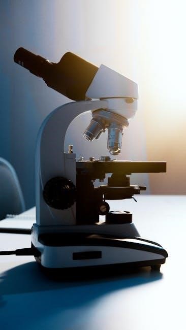
compound light microscope parts and functions worksheet pdf
The compound light microscope is a fundamental tool in biology and medicine, enabling magnification of specimens for detailed observation. It uses a combination of lenses and light to produce enlarged images, advancing scientific research and education. Understanding its parts and functions is essential for effective use in laboratory settings.
1.1 Overview of the Compound Light Microscope

A compound light microscope is an optical instrument that uses two lens systems to magnify specimens significantly. It is widely used in educational and research settings to study small structures. The microscope relies on visible light to illuminate samples, offering a detailed view of microscopic organisms and tissues. Its design includes essential components such as eyepieces, objective lenses, a stage, and light-control mechanisms. Worksheets and guides are often utilized to help users identify and understand its parts and functions, making it an indispensable tool for learning microscopy basics and conducting scientific investigations effectively.
1.2 Importance of Understanding Microscope Parts and Functions
Understanding the parts and functions of a compound light microscope is crucial for its effective use. Proper knowledge ensures accurate specimen observation, preventing damage to both the microscope and samples. It enhances experimental outcomes by allowing precise adjustments for optimal image clarity. Additionally, comprehension of components like the eyepiece, objective lenses, and condenser fosters better maintenance and troubleshooting. Worksheets and guides are essential tools for mastering these concepts, making them invaluable for students and researchers alike. This foundational knowledge is key to unlocking the full potential of microscopy in scientific exploration and education.

Key Parts of the Compound Light Microscope
The compound light microscope consists of essential components like the eyepiece, objective lenses, revolving nosepiece, stage, and stage clips. Each part plays a vital role in achieving clear images.
2.1 Eyepiece (Ocular Lens)
The eyepiece, or ocular lens, is a critical component of the compound light microscope. Positioned at the top of the microscope, it serves as the lens through which the user views the magnified image. The eyepiece works in conjunction with the objective lens to further magnify the specimen. Typically, eyepieces have a magnification power of 10x or 15x. They are designed to focus the image formed by the objective lens, ensuring clarity and detail. Proper adjustment of the eyepiece is essential for comfortable viewing, especially for users with vision corrections like astigmatism or nearsightedness.
2.2 Objective Lenses

The objective lenses are a set of high-powered lenses located near the specimen stage of the compound microscope. They are mounted on a revolving nosepiece, allowing easy switching between different magnifications. Objective lenses are responsible for focusing light from the specimen and forming the initial magnified image. Common magnifications include 4x, 10x, 40x, and 100x (oil immersion). Each lens is designed for specific viewing needs, with higher magnification lenses requiring more precise focus and light adjustment. Proper alignment and cleaning of objective lenses are essential for optimal image clarity and microscope performance.
2.3 Revolving Nosepiece
The revolving nosepiece is a circular platform that holds multiple objective lenses of different magnifications. It is designed to rotate smoothly, allowing quick and easy switching between lenses without disturbing the specimen. This feature enhances efficiency in observing specimens at varying magnifications. The nosepiece typically includes click-stops to ensure precise alignment of the selected lens with the eyepiece. Proper alignment is crucial for maintaining focus and image clarity when changing lenses. The revolving nosepiece is a key component that simplifies the process of adjusting magnification during microscopy, making it an essential part of the microscope’s functional design.
2.4 Stage and Stage Clips

The stage is a flat platform located below the objective lenses, designed to hold the specimen in place for observation. It is typically equipped with stage clips or clamps that securely fasten the microscope slide, ensuring the specimen remains steady during examination. The stage often includes a mechanical stage that allows for precise movement of the slide, enabling the user to focus on different areas of the specimen. Properly securing the slide with stage clips is crucial for clear and accurate viewing under magnification, as any movement could blur the image or misalign the specimen.

Functions of the Microscope Parts
The microscope parts work together to magnify specimens, focusing light through lenses to produce clear images. Each component has a specific role in enhancing visibility and precision.
3.1 Magnification and Image Formation
Magnification in a compound light microscope occurs through two lens systems: the eyepiece and the objective lens. The eyepiece acts as a magnifying glass, while the objective lens focuses light from the specimen; Together, they create a larger virtual image. Total magnification is the product of the eyepiece and objective lens powers. Proper alignment of light and precise focus are critical for clear image formation. The microscope’s ability to produce detailed, enlarged images makes it indispensable in scientific exploration and education. Understanding this process enhances the ability to use the microscope effectively for observing microscopic structures.
3.2 Focusing Mechanisms (Coarse and Fine Adjustment Knobs)
The coarse adjustment knob moves the stage up or down for initial focusing, bringing the specimen into approximate view. The fine adjustment knob makes precise, minor adjustments to achieve sharp focus. Together, they ensure clear, detailed observation of specimens. Proper use of these knobs is essential for efficient microscopy, preventing damage to the slide or objective lens. Mastering their operation enhances the accuracy of microscopic examinations in educational, research, and diagnostic settings.
3.3 Light Control (Condenser and Diaphragm)
The condenser focuses light onto the specimen, enhancing image clarity and contrast, while the diaphragm regulates the amount of light entering the microscope; The diaphragm, located below the stage, has apertures of varying sizes to control light intensity; Proper adjustment ensures optimal illumination, preventing overexposure or underexposure of the specimen; Together, these components are essential for achieving high-quality images in microscopy, allowing for precise observations in scientific and educational settings. Their correct use is vital for maximizing the effectiveness of the compound light microscope.

Worksheet Activities for Learning Microscope Parts
Worksheet activities, such as labeling diagrams and matching parts to their functions, enhance understanding of microscope components. These exercises are essential for hands-on learning and retention.
4.1 Labeling Diagrams
Labeling diagrams are a cornerstone of learning microscope parts, enhancing visual recognition and understanding. Worksheets often feature detailed illustrations of microscopes, requiring students to identify and label components like the eyepiece, objective lenses, stage, and condenser. This interactive approach aids in memorizing the structure and functions, fostering a deeper connection with the material. By associating names with visual elements, learners build a foundational knowledge crucial for practical applications. These activities are particularly effective in classroom settings, making complex concepts accessible and engaging for students of all levels. Accurate labeling ensures clarity and precision, essential skills for future scientific endeavors.
4.2 Matching Parts to Their Functions
Matching parts to their functions is an interactive way to reinforce understanding of microscope components. Worksheets often present lists of parts, such as the eyepiece, objective lenses, and condenser, alongside their roles. Students link each part to its function, fostering a clear grasp of how each element contributes to the microscope’s operation. This activity strengthens memory retention and logical thinking, as learners connect visual components with their practical uses. By mastering this skill, students gain confidence in using microscopes effectively, preparing them for hands-on laboratory work and advanced scientific exploration. This method ensures a comprehensive understanding of the microscope’s functional design and purpose.
Mastering the compound light microscope’s parts and functions is crucial for scientific exploration. Additional resources, including worksheets and guides, provide further learning opportunities for students and researchers.
5.1 Summary of Key Concepts
The compound light microscope is a vital tool for magnifying small specimens, combining eyepiece and objective lenses for enhanced observation. Key components include the eyepiece, objective lenses, revolving nosepiece, stage, and focus knobs, each serving distinct roles in image formation and clarity. The microscope’s functionality relies on light control via the condenser and diaphragm, ensuring optimal illumination. Understanding these parts and their functions is crucial for effective microscopy, advancing both educational and research applications. This guide provides a comprehensive overview, enabling users to master microscope operations and explore the microscopic world with precision and confidence.
5.2 Additional Resources for Further Learning
For deeper understanding, explore online guides and educational websites offering detailed explanations of microscope parts and functions. Downloadable worksheets and interactive modules provide hands-on practice, reinforcing learning. Science educational platforms feature labeled diagrams, quizzes, and activities to test knowledge. Additionally, academic resources such as PDF manuals and tutorials offer comprehensive insights into microscopy techniques. These tools are ideal for students and educators seeking to enhance their skills in microscopy, ensuring a solid foundation for future scientific exploration and experimentation.